Difference between sonography & colour doppler ultrasound is pretty narrow, because the basic technology used to perform this test on the patient is almost same. That is ultrasound!
There are different types of ultrasound imaging, including sonography and colour Doppler ultrasound. While both methods use high-frequency sound waves to create images of the inside of the body, there are some key differences between the two.
So, here in this article, we going to explain to you the fundamental difference between normal sonography & advanced colour Doppler.
So, first of all, let’s see what is Sonography(3D-4D Sonography).
Sonography, also known as ultrasound sonography, is a medical imaging technique that uses high-frequency sound waves to produce images of the body’s internal organs and tissues. It is commonly used to visualize the uterus, ovaries, and other reproductive organs, as well as the liver, kidneys, and other organs.
Sonography is a non-invasive, painless, and safe procedure that does not involve the use of radiation. It is often used as an alternative to X-ray or CT (computed tomography) scans, which use ionizing radiation.

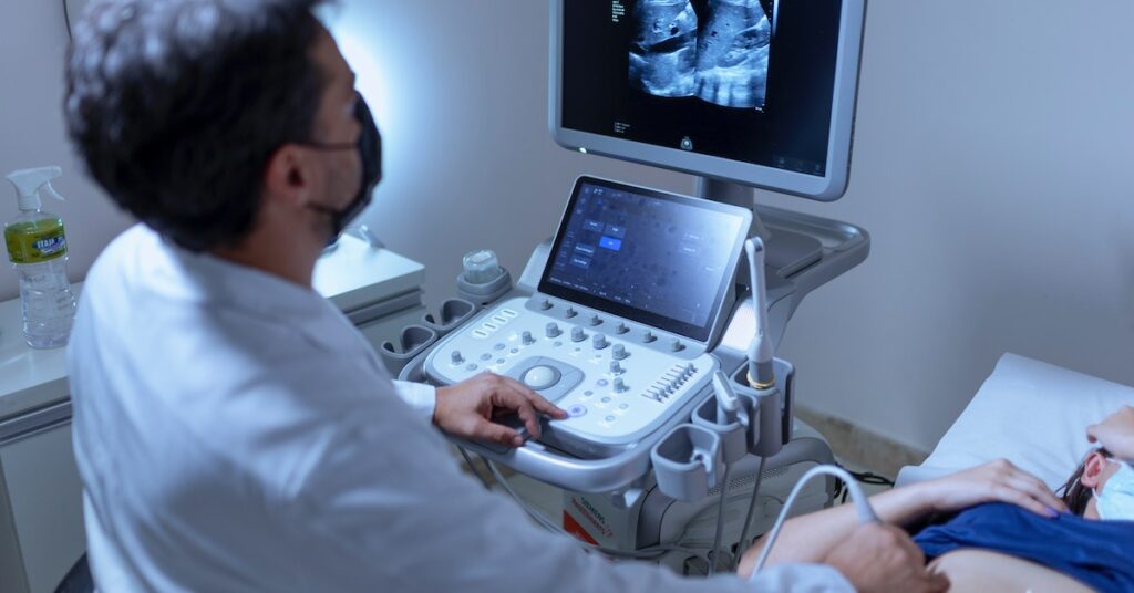
To perform a sonography procedure, a healthcare provider will place a small device called a transducer on the patient’s skin. The transducer sends and receives high-frequency sound waves, which bounce off of the organs and tissues inside the body.
The reflected sound waves are then detected by the transducer and sent to a computer, which processes the data and creates images of the organs and tissues.
Sonography is typically used to evaluate the health and function of the organs and tissues, to detect abnormalities or conditions such as tumours, cysts, or infections, and to monitor the development of fetuses during pregnancy.
Now, let’s see What Is Colour Doppler Ultrasound?
Colour Doppler ultrasound is a medical imaging technique that uses ultrasound to produce images of the body’s internal organs and tissues, while also providing information about the flow of blood through the vessels.
Similar to sonography, during the procedure, a healthcare provider will place a small device called a transducer on the patient’s skin. The transducer sends and receives high-frequency sound waves, which are used to create images of the organ or tissue being examined.
The sound waves also bounce off of moving red blood cells, allowing the healthcare provider to see the direction and velocity of blood flow. Colour Doppler ultrasound is commonly used to evaluate the blood flow in the arteries and veins, as well as to assess the function of the heart, liver, kidneys, and other organs. It can be used to diagnose conditions such as blockages in the arteries, blood clots, and abnormalities in the heart’s structure.
In the images produced by colour Doppler ultrasound, the flow of blood is represented by different colours, with red indicating blood flowing towards the transducer and blue indicating blood flowing away from the transducer. The intensity of the colour corresponds to the velocity of the blood flow, with brighter colours indicating faster flow.
Wrapping it Up
These were the main difference between the two ultrasound technologies used for evaluation & different purposes. The colour doppler ultrasound is very useful in the healthcare industry, helping to identify any issue upfront.
3D-4D Sonography in Ahmedabad
Infocus Diagnostics is a chain of advanced diagnostic centres located all over Ahmedabad, Gujarat, with extensive diagnosis facilities under one roof. We are integrated with modern infrastructure, advanced technology and specialised expertise to provide colour doppler ultrasound & sonography. Book your appointment to get the best diagnosis for a Doppler Test in Ahmedabad.