Ultrasound of the heart is commonly called an “echocardiogram” or “echo” for short.
A 2D echocardiogram provides your physician with information like the functioning of your heart, diagnosing the malfunctions, if any, and planning the treatment for the developing disease.
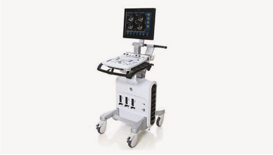
During a 2D echocardiography procedure, a healthcare provider will place a small transducer on the patient’s chest. The transducer sends and receives high-frequency sound waves, which create an image of the heart utilising an advanced 2D echo machine. The images are displayed on a screen, allowing the healthcare provider to evaluate the heart’s size, shape, and function.
Doctors may recommend it to:
No. It’s a safe, painless procedure with no radiation exposure. It only involves a probe being moved over your chest with a gel.
No special preparation is needed. You can eat and take your medications as usual, unless advised otherwise by your doctor.
Stay connected with Infocus Diagnostics for expert health tips, exclusive offers, and the latest updates in diagnostic care. Join our growing community and be the first to know how we’re making healthcare more accessible, accurate, and patient-focused — one post at a time.
Radiology – 7969027277
Pathology – 9099088948
helpdesk@infocusdiagnostics.com
Navrangpura 45/B, Swastik Society, Opp. Vipul Dudhiya, Stadium-Commerce Six Roads, Navrangpura, Ahmedabad-380009.