3 T MRI (TIM+DOT)
What is an MRI ?
MRI (magnetic resonance imaging) is a medical imaging technique that uses strong magnetic fields and radio waves to create detailed images of the body’s internal structures. It is a non-invasive and painless procedure that is used to diagnose and monitor various medical conditions, including cancer, cardiovascular disease, neurological disorders, and musculoskeletal injuries. MRI can also be used to guide surgical procedures and to monitor the effectiveness of treatments.
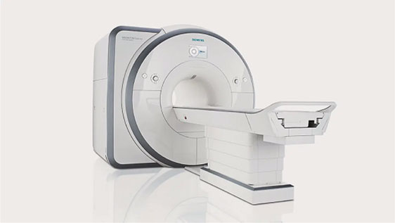
What is a 3T MRI ?
A 3T MRI (3 Tesla MRI) is a type of MRI machine that uses a stronger magnetic field than standard MRI machines. The “Tesla” unit refers to the strength of the magnetic field, and a 3T MRI has a magnetic field that is three times stronger than a standard 1.5T MRI. This stronger magnetic field allows for higher resolution images and greater accuracy in diagnosing certain medical conditions. 3T MRI machines are often used for brain imaging and are particularly useful for detecting small abnormalities or changes in the brain tissue. However, they may not be suitable for all patients, such as those with certain types of implants or devices, and may require special precautions to be taken before the scan.
Advantages of 3T MRI
3T MRI offers several benefits, including the ability to produce highly detailed and accurate images of the body’s internal structures. It also has a faster imaging time and is able to capture high-quality images of soft tissues and vital organs. Additionally, 3T MRI machines have a larger bore or tube, allowing patients to keep their head outside during certain scans. While 3T MRI is generally safe and does not have serious side effects, some mild, temporary side effects may occur.
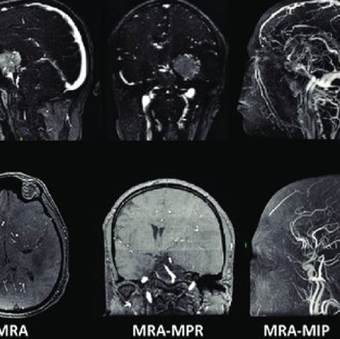
Neuro Imaging
Neuroimaging is used to diagnose disease and assess brain health. Neuroimaging also studies how the brain works and how various activities impact the brain.
Neuro Imaging Services
- Conventional Brain and Spine Imaging
- MR Angio and MR Venography
- Diffusion Tensor Imaging
- Brain Perfusion
- Multivoxal Spectroscopy
- Propeller Imaging
- MERGE & COSMIC Spine Imaging
- CSF Cisternography
Musculoskeletal Imaging
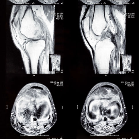
Musculoskeletal Imaging involves viewing- interpreting medical images of bones, joints and related soft tissues and diagnosing injuries and disease.
Musculoskeletal Imaging Services
- All joints scan
- Cartilage evaluation with 3D DESS
(Dual Echo study state gradient recalled echo)
Abdominal Imaging
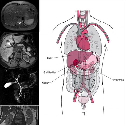
Abdominal magnetic resonance imaging (MRI) is a noninvasive procedure that uses powerful magnets and radio waves to produce pictures of the inside of the abdomen without exposure to ionizing radiation (x-rays).
Abdominal Magnetic Services
- Conventional Abdomen- Pelvis Imaging
- MRCP
- Liver Imaging (Triphasic Scan)
- MR Defecography
- MR Enterography
- MR Urography
- Renal Angiography
- MP MRI for Prostate
- Male/Female Pelvic MRI
- Abdominal Aortogram
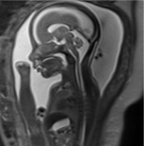
FETAL MRI
A fetal MRI is used as an additional tool for advanced evaluation of structural abnormalities especially involving the fetal brain, bowel, head/neck tumours, spine, lungs and placenta. It has been proven to be safe for use in the 2nd and 3rd trimester of pregnancy and has no radiation exposure.
We are the only centre in Gujarat and among the few in India to provide expert MRI scans of a fetus with an experience of more than 10 years.
![]()
Click Here To Know More About Preparations & Procedure
BREAST MRI
Magnetic resonance imaging (MRI) of the breast or breast MRI is a test used to detect breast cancer and other abnormalities in the breast. A breast MRI may be used with mammograms as a screening tool for detecting breast cancer. That group of people includes women with a high risk of breast cancer, who have a very strong family history of breast cancer or carry a hereditary breast cancer gene mutation.
Indications for Breast MRI
- Diagnosed breast cancer for the extent of disease.
- A suspected leak or rupture of a breast implant.
- High risk for breast cancer, defined as a lifetime risk of 20% or greater, as calculated by risk tools that account for family history and other risk factors.
- A strong family history of breast cancer or ovarian cancer.
- Very dense breast tissue or mammograms findings are difficult to diagnose.
- A history of precancerous breast changes — such as atypical hyperplasia or lobular carcinoma in situ — and a strong family history of breast cancer and dense breast tissue.
- Hereditary breast cancer gene mutation, such as BRCA1 or BRCA2.
- Prior radiation treatments to the chest area before age 30 years.
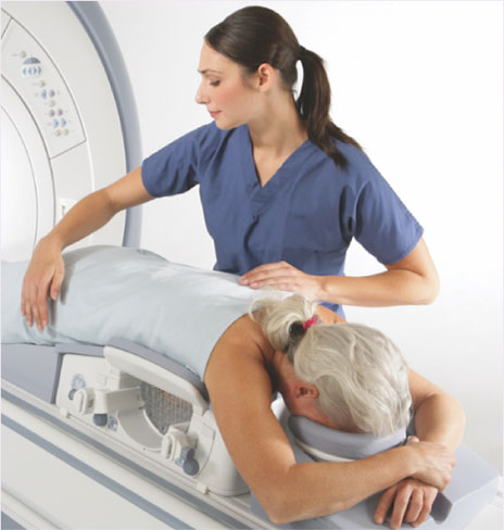

3T MRI Services Preparations & Procedures
FETAL MRI Details
A fetal MRI is a specialized imaging test that may be helpful in evaluating your fetus for congenital anomalies and associated complications. Like an ultrasound, an MRI does not use the type of radiation that can cause DNA damage, so we believe it is safe to perform in pregnancy
MRI cannot be done in patients with
1. Metallic & electronic objects inside your body.
2. Pacemaker, Cochlear Implant, and prosthesis.
3. Bullets or shrapnel.
4. In 1 st trimester of pregnancy.
Instructions before MRI Scan
1. Before entering the scan room all jewelry items, Metal things such as hair clips, jewelry,
watches, belt, credit cards, coins, hearing aids, and dentures should be removed.
2. Bring all your previous files, reports & current medications list if there.
3. Patient or relative should inform the technician if a patient is claustrophobic or having
anxiety issues.
After MRI scan
- You can resume your routine activities after the MRI scan unless you were given a sedative
